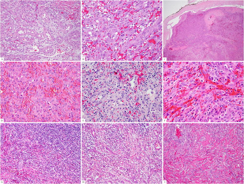
Figure 1. Pathologic findings of the FOS-rearranged EH cases.
(A) The index case (case #1), positive for FOS-LMNA fusion, showed a typical morphology with epithelioid cells surrounding vascular lumina. (B) Lesional cells showed glassy eosinophilic cytoplasm with scattered vacuoles, vesicular nuclei, and prominent nucleoli (case #1). (C) One of the cellular EH (case #14) was confined to a dilated vascular channel, which at higher power (D) was composed of solid sheets of tumor cells. Cellular EH showed (E) variably prominent vascular spaces (case #12), (F) distinctive epithelioid cell features (case #11), and (G) surrounding lymphoid and eosinophilic infiltrates (case #13). (H) One typical EH was characterized by a prominent inflammatory background obscuring the proliferating vascular channels (H, case #15). Eosinophils were usually seen. (I) The penile EH (case #4) was composed of typical vasoformative structures but showed a more infiltrating growth pattern.

