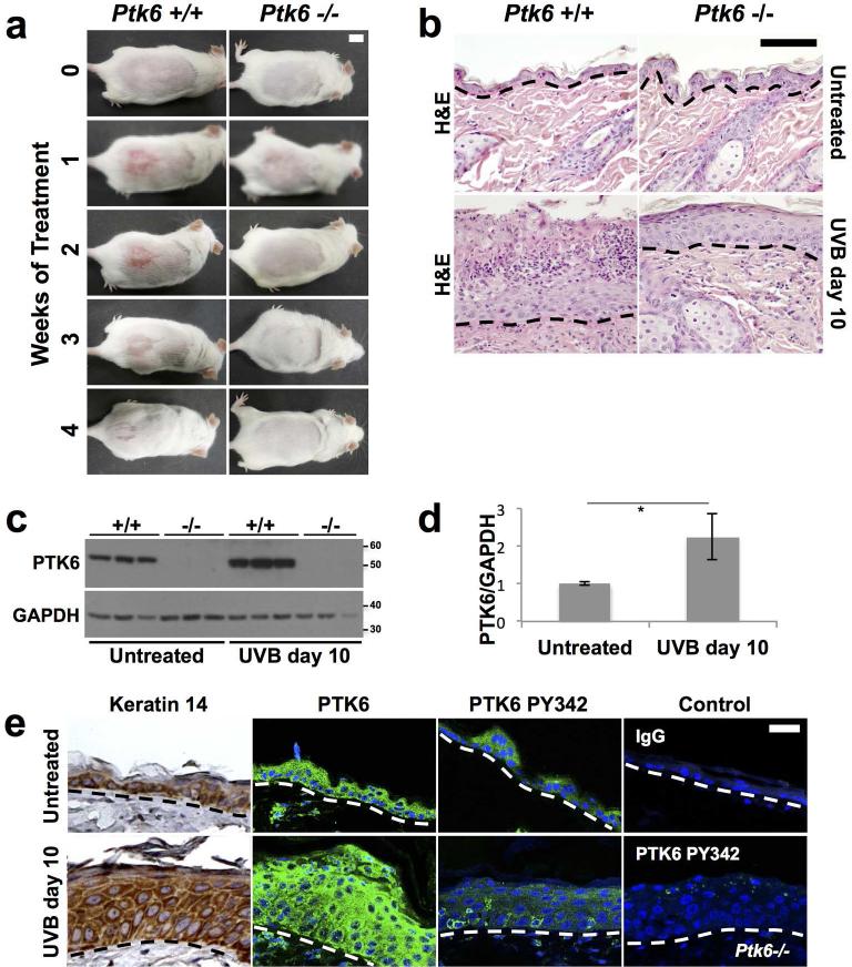
Figure 2. Increased Inflammation and PTK6 expression are detected in wild type mice after UVB treatment.
A: Increased and prolonged inflammation was detected in wild type mice compared with Ptk6−/− mice after short term exposure to UVB. Ptk6+/+ mice (left panels) developed erythema and an inflammatory reaction on the lower dorsal skin within a week after beginning UVB treatments. Mice were treated with UVB three times a week. Size bar = 1 cm. B: Increased degeneration and necrosis of the upper epidermal layers with neutrophilic migration and microabscess formation was detected in wild type mice when compared with Ptk6−/− skin at 10 days post initiation of short term UVB treatment. Representative H & E stained sections are shown. C: PTK6 expression is induced by UVB treatment in mouse skin. Lysates of adult (8-weeks old) mouse skin after short-term UVB were prepared and analyzed by immunoblotting. Each lane represents a sample from a different mouse. GAPDH was used as a loading control. D: The increase in PTK6 expression after UVB treatment (2C) was quantified using ImageJ (Rasband, 2011). The intensity of PTK6 signal was normalized to intensity of the GAPDH loading control and averaged across all samples. p-value = 0.025. E: Total PTK6 and active PTK6 Y342 expression was examined by immunofluorescence in untreated and UVB-treated skin. Keratin 14 is expressed throughout the hyperplastic skin after UVB treatment. Controls included staining with IgG and staining of UVB-treated Ptk6−/− skin with the antibody against PTK6 PY342. No specific signal for PY342 was detected in Ptk6−/− mice. Size bar = 20 μm.

