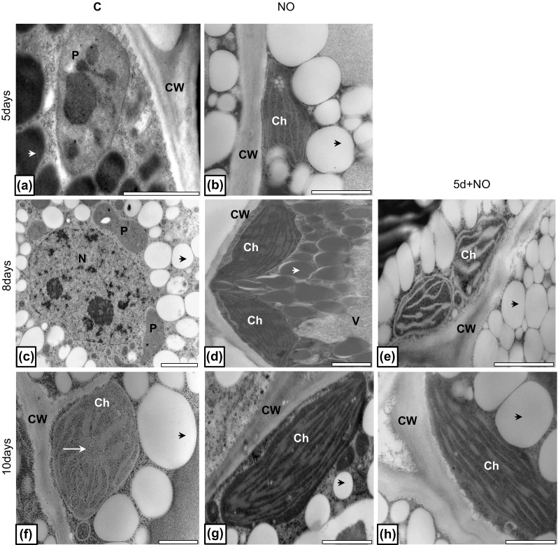
Fig. 8.

Ultrastructure of cells of upper cotyledon of control (a, c, f), NO-treated embryos (b, d, g) and plantlets developed from abnormal embryos treated with NO after 5 days of growth (e, h). Photographs were made after 5 (a, b), 8 (c, d, e) and 10 (f, g, h) days of culture. Micrographs are representative for ultrastructure of upper cotyledons of embryos after each treatment and specified period of culture. Black arrow cytoplasmic domain rich in lipid bodies; white short arrow cytoplasmic protein bodies; white long arrow prolamellar body, CW cell wall, P proplastid, Ch chloroplast, V vacuole. Bar 2 µm (a, c–e, g), 1 µm (b, f, h)
