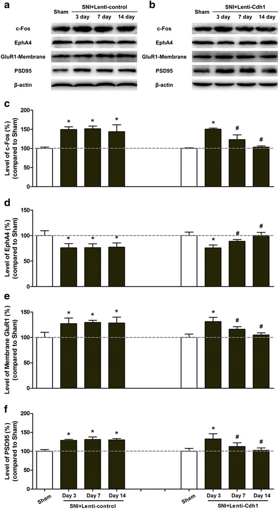
Fig. 5.

Intra-ACC microinjection of Cdh1-expressing lentivirus normalised SNI-induced redistribution of AMPA receptor GluR1 subunit in ACC. a, b Representative Western blotting of c-Fos, EphA4, membrane-bound GluR1, and PSD-95 in the ACC of rats subjected to SNI 3, 7, and 14 days following intra-ACC microinjection of control lentivirus (Lenti-control, a) or Cdh1-expressing lentivirus (Lenti-Cdh1, b). c Pooled data showing that Cdh1-expressing lentivirus markedly reduced the SNI-induced increase in c-Fos expression in the ACC 7 and 14 days after lentivirus microinjection (n = 3). Error bars SD; *p < 0.05 vs. Sham; # p < 0.05 vs. control lentivirus. d Microinjection of Lenti-Cdh1 into the ACC significantly increased EphA4 levels 7 and 14 days after lentivirus injection (n = 3 per group). Error bars SD; *p < 0.05 vs. Sham; # p < 0.05 vs. control lentivirus. e In the ACC, the SNI-induced increase in cell surface GluR1 expression was gradually returned to control levels 7 and 14 days after microinjection of Lenti-Cdh1. Results are expressed as mean ± SD (n = 3, each group); *p < 0.05 vs. Sham; # p < 0.05 vs. Lenti-control. f The SNI-induced increase in PSD-95 protein expression in the ACC was gradually normalised 7 and 14 days after Lenti-Cdh1 injection. Results are expressed as mean ± SD (n = 3); *p < 0.05 vs. Sham; # p < 0.05 vs. Lenti-control
