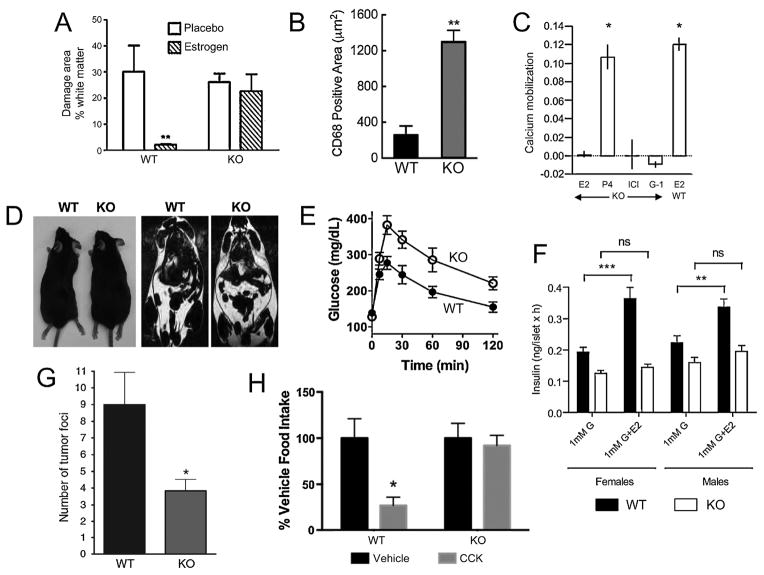
Figure 1.
GPER deficiency leads to multiple phenotypes and loss of estrogen responsiveness of multiple tissues. A. The estrogen-mediated decrease in white matter damage, a result of immune infiltration and demyelination, in a murine model of multiple sclerosis (experimental autoimmune encephalomyelitis, EAE) is absent in GPER KO1 mice [98]. B. GPER KO1 mice fed an atherogenic diet show increased vascular infiltration of macrophages (CD68-positive cells) associated increased atherosclerosis [124]. C. Renal intercalated cells from GPER KO2 mice are deficient in estrogen (E2)-, ICI182,780 (ICI)- and G-1-mediated calcium mobilization. Calcium mobilization to progesterone (P4) was unaltered in GPER KO2 mice, similar in magnitude to estrogen-mediated calcium mobilization in WT cells [132]. D. GPER KO1 mice exhibit increased body size/obesity (left) and adiposity (right, as measured by MRI) [101]. E. Upon aging, male GPER KO1 become glucose intolerant [101]. F. Estrogen (E2)-mediated insulin secretion from islets is absent in both male and female GPER KO2 mice [50]. G, glucose. G. GPER KO1 mice exhibit reduced metastasis in a PyMT-MMTV model of mammary tumorigenesis [160]. H. Female GPER KO1 mice display a lack of cholecystokinin (CCK)-mediated satiety/appetite suppression (an estrogen-sensitized effect) as measured by food intake [139]. Reproduced (in some cases with minor modifications for clarity) with permission from the indicated sources.

