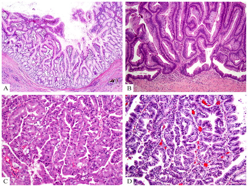
Figure 5.
A) Gastric type IPMN shows relatively simple and typically short papillae and often have pyloric-like glandular elements at their base in the cyst wall. The epithelial lining is highly similar to gastric foveolar epithelium. B) Intestinal type IPMN typically has a villous growth pattern and reveals pseudostratified columnar cells with a basophilic appearance and apical mucin. C) Oncocytic type IPMN reveals arborizing papillae lined by 2–5 layers of cuboidal cells with oncocytic cytoplasm and prominent, eccentric nucleoli as well as intraepithelial lumina. D) Pancreatobiliary type IPMN has complex arborizing and interconnecting papillary configurations with delicate fibrovascular cores and is composed of cuboidal cells with enlarged nuclei and little mucin production (hematoxylin and eosin stain).

