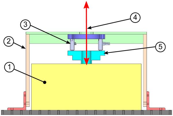
Figure 3.

Schematic section view of the experimental setup. 1) Location of where the Ecoflex sample or human forearm is placed, rectangle represents a simple section view of an Ecoflex sample. 2) Al 6061 box frame. 3) Three preloaded piezos. 4) SLDV LASER. 5) Annular mechanical radiator. Not shown is the SLDV head located approximately 300 mm above the sample surface and the third piezo.
