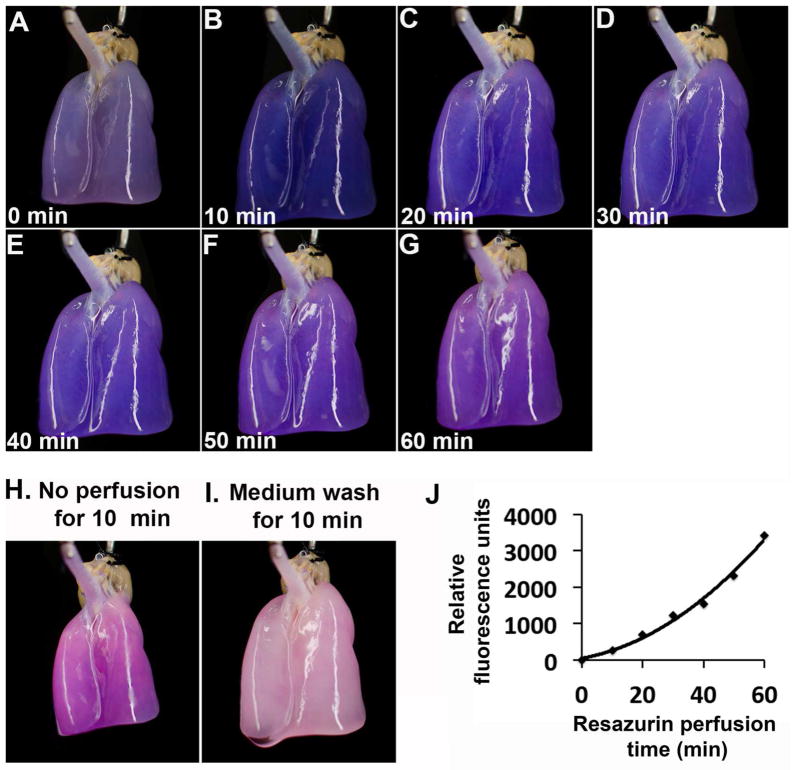
Fig. 2.
Resazurin perfusion of a HUVEC-regenerated lung. (A–G) Time-lapse photography of the HUVEC-regenerated lung during resazurin perfusion. Images were taken at 0, 10, 20, 30, 40, 50 and 60 min after the start of resazurin perfusion. (H) An image was taken after the resazurin perfusion was paused for 10 min. (I) A final image was taken after the resazurin-containing medium was replaced with fresh medium without resazurin and perfused the lung for an additional 10 min. (J) Fluorescence intensities of medium sampled at each 10 min interval during the resazurin perfusion.

