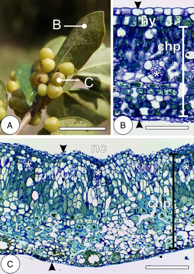
Figure 1.

Morphology and anatomy of Psidium cattleianum leaves and Nothotrioza myrtoidis galls. (A) The detail of a simple leaf with globoid galls protruded to the abaxial surface. (B) Cross-section of mature leaf with uniseriate epidermis on both surfaces (arrowheads), hypodermis (hy) and vascular bundles (asterisks) interspaced to the dorsiventral chlorophyllous parenchyma (chp). (C) Cross-section of mature gall with uniseriate epidermis on both surfaces (arrowheads) and vascular bundles (asterisks) near the nymphal chamber (nc) interspaced to the homogenous chlorophyllous parenchyma (chp). Bars: (A) 3 cm; (B) 100 µm; (C) 200 µm.
