Figure 2.
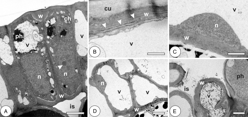
Cytology of epidermal cells in the leaves of Psidium cattleianum and galls of Nothotrioza cattleiani. (A–C) Leaves. (D and E) Galls. (A) Cells with thin anticlinal primary walls (w), large nuclei (n) with conspicuous nucleoli (arrowhead), few chloroplasts (ch) and small vacuoles (v), with phenolic inclusions (ph). (B) The detail of a cell with polylamellate (arrowheads) cell wall (w), thick cuticle (cu) and hyaline vacuole (v). (C) The detail of a cell with homogenous secondary wall (w), small nucleus (n) and hyaline vacuole (v). (D) Induction phase. Cells with homogenous thickened walls (w), peripheral nuclei (n) and hyaline vacuoles (v). Discrete sites of periclinal divisions are observed (asterisk). (E) Maturation phase. Intermittent cell layer, with intercellular spaces (is), heterogeneous thickened and polylamellate walls and inconspicuous cuticle. Vacuoles (v) may contain phenolic inclusions (ph). Bars: (A–C) 2 µm; (D and E) 5 µm.

