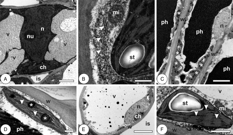
Figure 3.
Cytology of chlorophyllous parenchyma cells in leaves of Psidium cattleianum. (A and B) Young leaves. (C–F) Mature leaves. (A) Cell with primary thin walls, large central nucleus (n) with conspicuous nucleolus (nu), fragmented vacuole (v) and chloroplasts (ch) with well-developed grana. Intercellular spaces (is) are reduced. (B) The detail of a cell with dense cytoplasm, RER (arrowheads) and mitochondria (mi) associated with chloroplasts with well-developed grana and starch grain (st). (C) Palisade parenchyma cells with homogenous walls (w), phenolic-rich vacuoles (ph), many chloroplasts with well-organized thylakoid lamellae and starch grains (asterisks) and peripheral nuclei (n) with inconspicuous nucleoli. (D) The detail of a chloroplast with low-stack grana (arrowheads) and large plastoglobules (asterisks). (E) Spongy parenchyma cell with homogenous primary wall (w), large vacuole (v), peripheral nucleus (n), chloroplasts (ch) and large intercellular space (is). (F) The detail of a chloroplast with well-developed thylakoid system, high-stack grana (arrowheads), starch grain (st) and associated mitochondria (mi). Bars: (A) 2 µm; (B and D) 500 nm; (C and E) 5 µm; (F) 1 µm.

