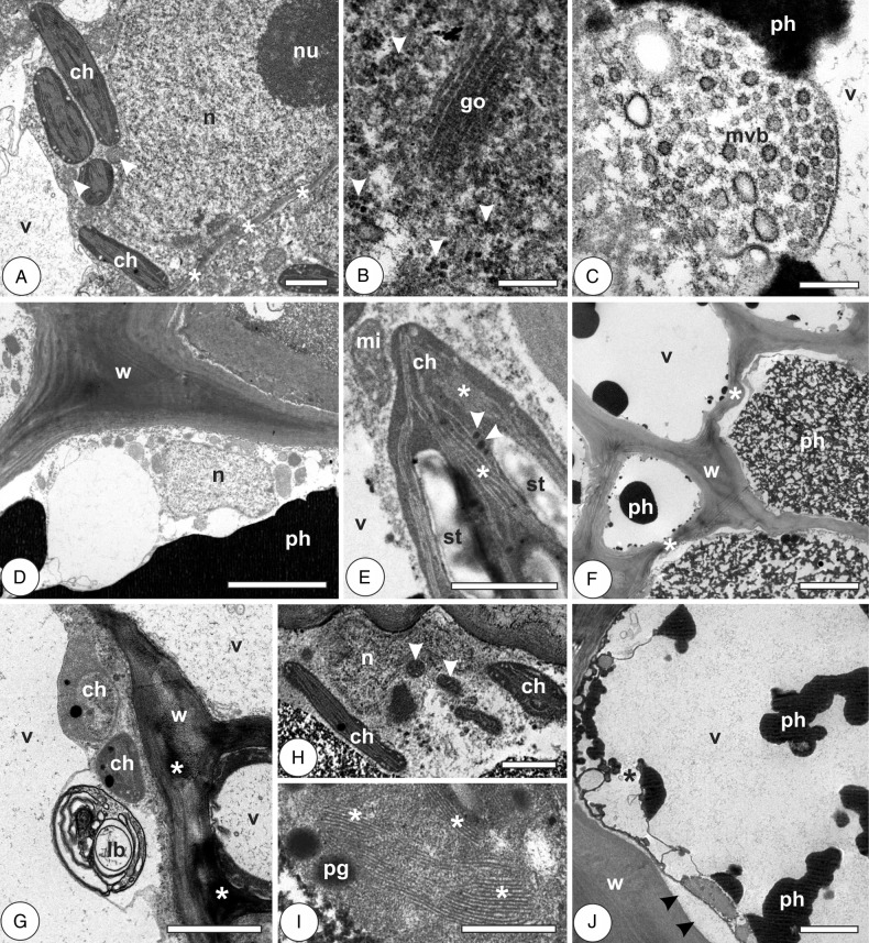
Figure 4.
Cytology of chlorophyllous parenchyma cells in the galls of Nothotrioza cattleiani. (A–C) Induction phase. (D and E) Growth and development phase. (F–H) Maturation phase. (I and J) Senescent phase. (A) The detail of a cell with large nuclei (n) with conspicuous nucleoli (nu), small mitochondria (arrowheads) associated with chloroplasts (ch) with well-organized lamellae. (B) Abundant polysomes (arrowheads) and Golgi apparatus (go). (C) Multivesicular body (mvb) at cytoplasm periphery. (D) Cells with secondary polylamellate walls (w), phenolic-rich vacuoles (ph) and small nuclei (n) with inconspicuous nucleoli. (E) Mitochondria (mi) associated to chloroplast (ch) with well-organized thylakoid lamellae (asterisks), starch grains (st) and small plastoglobules (arrowheads). (F) Cells with irregularly thickened walls (w, asterisks), phenolic (ph) or hyaline vacuoles (v). (G) The detail of lamellar body (lb), undeveloped chloroplasts (ch) and cell walls (w) with sites of different electron density (asterisks). (H) Degraded nucleus (n), small mitochondria (arrowheads) and chloroplasts. (I) Degraded chloroplast with vestigial grana (asterisks) and plastoglobules (pg). (J) Cell with large periplasmic spaces (arrowheads), disrupted vacuole (v, asterisk) with phenolic inclusions (ph). Bars: (A, E and H) 1 µm; (B and C) 200 nm; (D and F) 5 µm; (G and J) 2 µm; (I) 500 nm.

