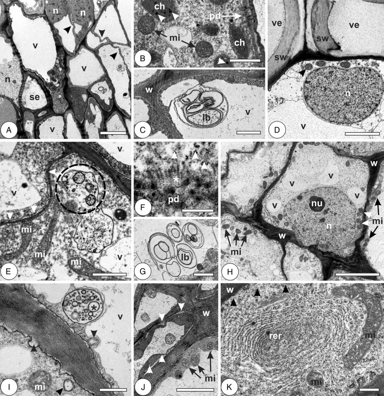
Figure 5.
Cytology of vascular and perivascular parenchyma cells in the leaves of Psidium cattleianum and galls of Nothotrioza cattleiani. (A–C) Young leaves. (D) Mature leaves. (E–K) Galls. (A) Cells with thin and sinuous walls (arrowheads), large nuclei (n) and hyaline vacuoles (v). Sieve elements (se) may be observed. (B) Cell with dense cytoplasm, many plasmodesmata (pd), round mitochondria (mi) and small underdeveloped chloroplasts (ch) with few small plastoglobules (arrowheads). (C) Lamellar body (lb) near the cell wall (w). (D) Cell with large nucleus (n) with dispersed heterochromatin, mitochondria (arrowhead) and hyaline vacuole (v). Vessel elements (ve) with secondary cell walls (sw) can be observed. (E–G) Induction phase. (E) Cells with thin sinuous walls (arrowheads), hyaline vacuoles (v), large mitochondria (mi) and multivesicular bodies (dashed circle). (F) The detail of plasmodesmata (pd) with aligned microtubules (asterisks) and abundant polysomes (arrowheads). (G) The detail of lamellar bodies (lb). (H and J) Growth and development phase. (H) Cells with irregularly thickened walls (w), fragmented hyaline vacuoles (v), large nuclei (n) with conspicuous nucleoli (nu) and abundant mitochondria (mi). (I) The detail of cells with multivesicular bodies (asterisk) and lomasomes (arrowheads). (J) Cells during maturation phase, with thick walls (w), abundant mitochondria (mi) and periplasmic phases (arrowheads). (K) Cell during senescent phase, with abundant rough endoplasmic reticulum (rer), polysomes (arrowheads) and large mitochondria (mi). Bars: (A and H) 5 µm; (B, C, E and G) 1 µm; (D and J) 2 µm; (F, I and K) 500 nm.

