Fig. 6.
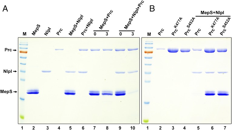
MepS is a substrate of Prc. (A) In vitro degradation assays. The C-terminal hexahistidine-tagged MepS, NlpI, and Prc proteins were mixed in all combinations (as indicated) and incubated for 3 h at 37 °C followed by SDS/PAGE and Coomassie blue staining. Each reaction contained the following (approximately): MepS, 15 μg; NlpI, 2 μg; and Prc, 4 μg. The mixtures in lanes 7 and 9 served as controls (0 h; no incubation). M is a protein marker with the following molecular size range (in kDa): 170, 130, 100, 70, 55, 40, 35, 25, 15, and 10. Purified MepS was always seen on SDS/PAGE as a doublet, and N-terminal sequencing confirmed that both bands belong to MepS itself. (B) Specificity of Prc. Two mutant derivatives of Prc (carrying K477A or S452A alterations at the active site residues) did not show any proteolytic activity (even when higher concentrations were used) against MepS in the presence of NlpI.

