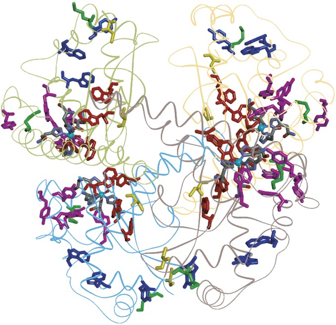
Fig. 4.
Structural model of the human SOD2 homotetramer [PDB ID 1N0J (33)]. Each polypeptide contains one five-coordinate Mn center (cyan ball and five ligands). Members of Tyr/Trp chains: red, 5-Å ET cutoff (Tyr34, Trp125, Trp123, and Trp161); magenta, 7.5-Å cutoff (Tyr11, Trp78, Tyr169, Tyr165, and Tyr176); blue, 10-Å cutoff (Trp181, Trp186, and Tyr193). Met residues are shown in green, and Cys residues are shown in yellow.

