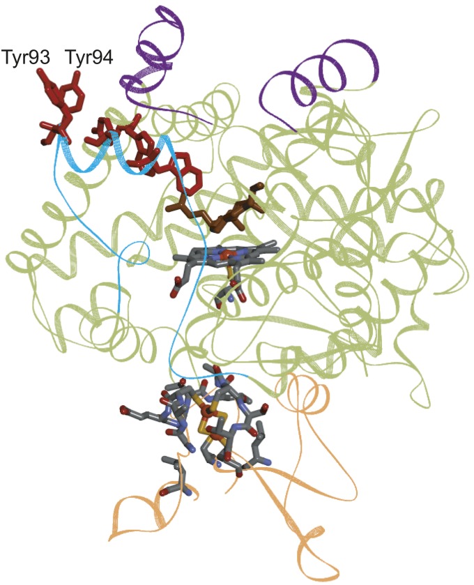
Fig. 5.
Structural model of human CYP11A1 (green ribbon) [PDB ID 3N9Y (38)] in complex with its redox partner adrenodoxin (orange ribbon). The purple ribbon highlights the inner mitochondrial membrane interaction zone, the cyan ribbon shows the hydrophilic water channel, and the cholesterol substrate is brown (38). The linear five-member Tyr/Trp chain is shown in red.

