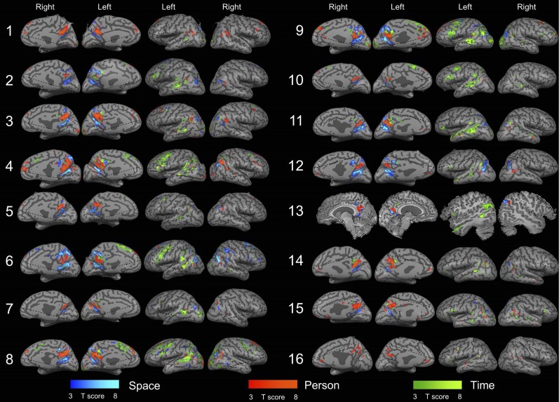
Fig. S1.
Cortical activity during orientation in space, time, and person in individual subjects. Domain-specific activity is shown for all 16 subjects, obtained by contrasting activity between each orientation domain and the other two domains (P < 0.05, FDR-corrected, cluster size >20 voxels). Dashed lines represent the limit of the scanned region in each subject. Subject 13 could not be transformed to an inflated brain representation due to technical reasons and is therefore presented by representative slices. Notice the consistent pattern of activity in the inferior parietal, medial parietal, frontal and temporal cortices.

