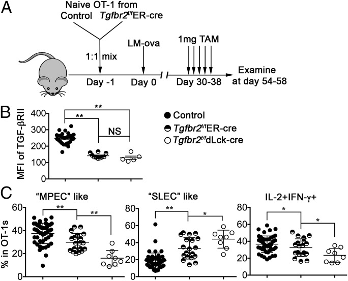
Fig. 1.
Delayed deletion of the TGF-β receptor alters memory T-cell population. (A) Schematic of the experiments. Naïve OT-1 T cells were purified from congenically marked control and Tgfbr2f/fER-cre mice and mixed at a 1:1 ratio. Then 104 mixed OT-1 cells were transferred into each B6 recipient mouse, followed by infection with 2,000 cfu of LM-ova administered i.v. on the second day. Starting at day 30 postinfection, 1 mg TAM or solvent alone was injected i.p. every other day for a total of five treatments. OT-1 T cells in the spleen were examined at the indicated times. (B) The mean fluorescence intensity (MFI) of TGF-βRII on OT-1 T cells was determined by flow cytometry. (C) Percentages of MPEC-like (Left), SLEC-like (Middle), and IL-2–producing cells (Right) within OT-1 T cells. Combined results from two independent experiments are shown. Each symbol in B and C represents data from an individual recipient mouse. **P < 0.01; *P < 0.05; NS, not significant, Student t test.

