Fig. 3.
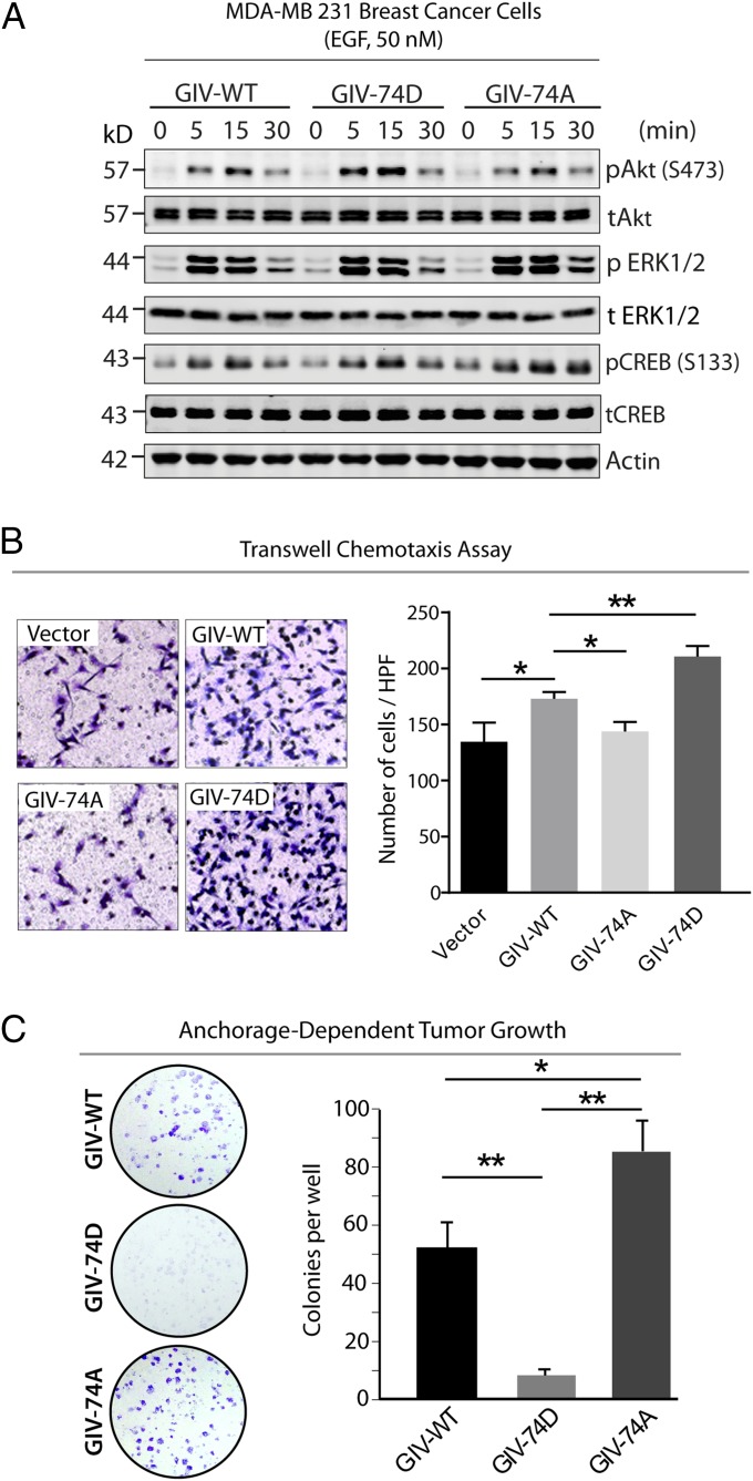
Phosphorylation of GIV at S1674 influences EGF signaling and influences preference for migration versus proliferation of MDA-MB231 cancer cells. (A) Serum-starved MDA-MB231 cells stably expressing GIV-WT or 74D/A mutants were stimulated with EGF before lysis. Whole-cell lysates were analyzed for total(t) and phospho(p) Akt, ERK, CREB, and actin by immunoblotting. (B) MDA-MB231 cells used in A were subjected to chemotaxis toward EGF, and migrating cells were visualized and analyzed as in Fig. 2D. Results are expressed as mean ± SEM; n = 3. HPF, high power field. (Magnification: 10×.) (C) MDA-MB231 cells used in A were analyzed for their ability to form adherent colonies on plastic plates for 2–3 wk before fixation and staining with crystal violet. (Left) Photograph of the crystal violet-stained wells. (Right) Bar graphs showing the number of colonies/cell line counted by ImageJ (colony counter).

