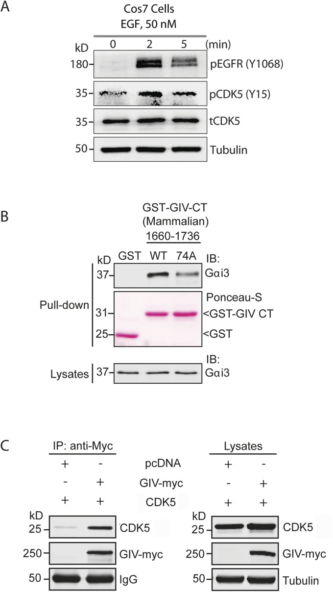
Fig. S5.
CDK5 is activated within minutes after EGF stimulation and binds and phosphorylates GIV. (A) EGF stimulation activates CDK5. Serum-starved Cos7 cells expressing CDK5 WT were stimulated with EGF before lysis. Whole-cell lysates were analyzed for phospho(pY1068)EGFR, total(t) and phospho(pY15) CDK5 and tubulin by immunoblotting. (B) Nonphosphorylatable GIV CT 1660–1736 mutant shows impaired binding to Gαi3 in cellulo. Cos7 cells were transiently transfected with WT or 74A mutant GST-GIV CT 1660–1736, and clarified cell lysates were incubated with glutathione-Sepharose beads. Bound proteins were analyzed by immunoblotting for endogenous Gαi3. Equal loading of GST and GST-GIV-CT proteins was confirmed by Ponceau-S staining (pulldown) and immunoblotting for Gαi3 (lysates). (C) CDK5 interacts with GIV. Cleared lysates obtained from Cos7 cells coexpressing pcDNA or GIV-myc and CDK5 were immunoprecipitated with anti-myc antibody. The immunoprecipitates were analyzed for bound CDK5 by immunoblotting with anti-CDK5 antibody. The expression of proteins was also checked by immunoblotting the lysates for the indicated proteins (Right).

