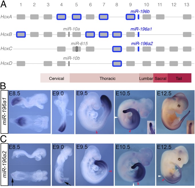
Fig. 1.
Unique and overlapping expression patterns of miR-196 paralogs in mouse. (A) Mouse Hox clusters, with the position of Hox-embedded microRNAs depicted. Predicted Hox targets of the miR-196 family are indicated in blue. (B and C) Detection of eGFP transcripts in miR-196a1GFP/+ (B) and miR-196a2GFP/+ (C) embryos demonstrates spatiotemporal expression differences for these identical miRNAs. Embryonic age indicated: red and white arrowheads indicate the anterior boundary of somitic and neural expression, respectively. A discrete band of reduced eGFP signal in the anterior PSM of later stage 196a1GFP/+ embryos is shown in Insets. Weak ventral eGFP signal in miR-196a2GFP/+ embryos at E8.5 and E9.0 is indicated with black arrows.

