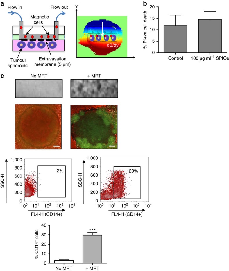
Figure 1. MRT using a novel transendothelial migration (TEM) flow assay.
We have designed a flow chamber that can accommodate three-dimensional (3D) tumour spheroids as well as a vascular endothelial layer. The flowing ‘magnetic cells' will therefore need to cross the vascular barrier before entering a 3D tumour simulating the passage of cells across endothelial cells in a blood vessel wall (a, left panel). The TEM flow chamber is placed in the isocentre of an MRI scanner with a spherical (6-mm diameter) homogenous 7 T magnetic field. We applied a pulsed gradient (50% of max) with strength of 300 mT m−1 in the (−y) gradient direction. The resulting heterogeneous magnetic field (dB/dy field) can steer magnetic particles towards the tumour spheroids for increased uptake (a, right panel). Cell viability following SPIO uptake was not compromised as assessed by flow cytometry for propidium iodide uptake (b). Uptake was confirmed by a distortion in the MRI image and a loss of signal compared with when no MRT was applied (c, upper panel). Corresponding fluorescent images of whole spheroids infiltrated with macrophages carrying a reporter adenovirus (Ad-CMV-GFP) are shown in (c, second panel). Scale bar, 100 μm. Flow cytometry of enzymatically dispersed spheroids revealed that the number of magnetic cells infiltrating spheroids (% of all cells present in spheroids that were CD14+) was significantly increased when a gradient was applied (c, lower panels). All images and data (means±s.e.m.) are derived from six independent experiments. Comparisons between the groups were performed using two-tailed unpaired Student's t-test ***P=0.0001, compared with no MRT group.

