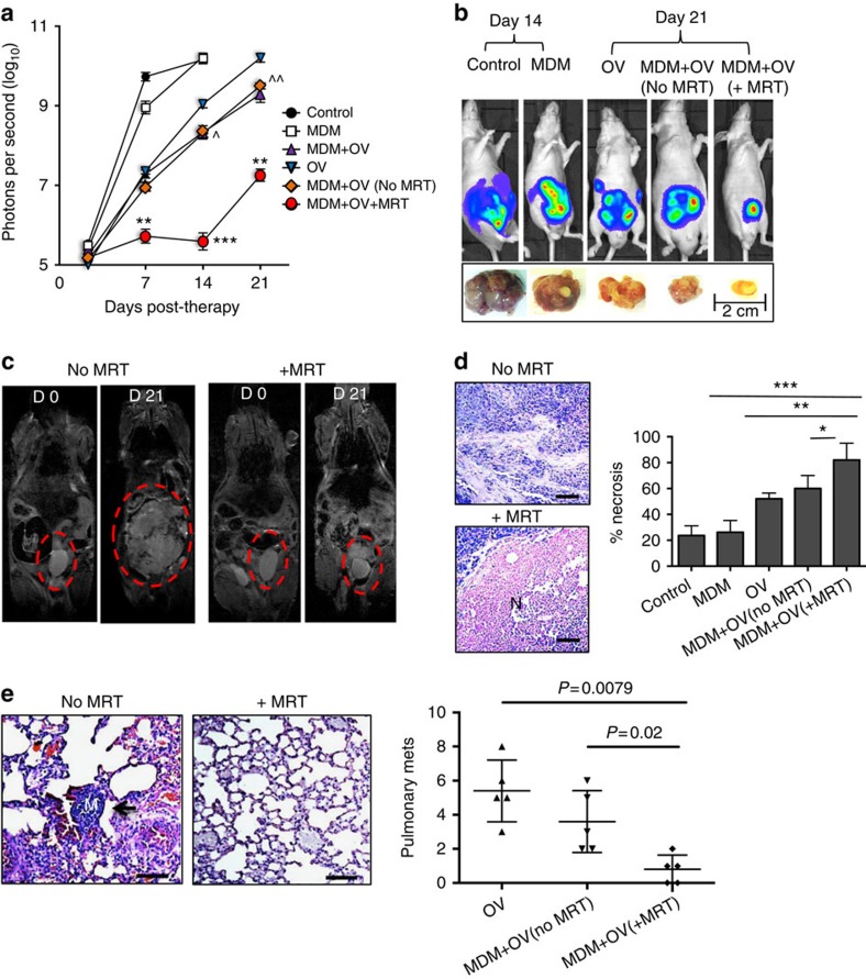
Figure 5. Magnetic targeting increases the anti-tumour effects of oncolytic macrophages.
Tumour-bearing mice were administered with a single dose of human monocyte-derived macrophages (MDMs) carrying the oncolytic virus, HSV1716 (MDM+OV). These were divided into three groups of mice (each with five mice per group). One group underwent MRT to either the prostate gland or lungs (MDM+OV+MRT) for 1 h, another was exposed to the MRI scanner but with no MRT (MDM+OV no MRT) and the third (MDM+OV) did not enter the MRI scanner. Additional groups of mice received 100 μl of PBS (Control), a single dose of 1 × 10⁷ plaque-forming unit HSV1716 (OV) or 3 million untreated MDM. Mice were imaged weekly using the IVIS imaging system and, after 21 days, tumours and lungs were removed and processed for histology. (a) Tumour luminosity in n=5 mice per group showed MRT significantly improved the effect of OV-MDM on tumour growth. (b) Representative IVIS images and photographs of primary tumours following various treatments. (c) Representative RARE images for MDM+OV with or without MRT show marked difference in tumour size at the beginning and end of therapy. Representative images of haematoxylin- and eosin-stained sections from n=5 mice per group show (d) the area of necrosis (N) in primary tumours and (e) the number of metastases (see arrow, M, metastasis) in the lungs of mice receiving MDM+OV with or without MRT. Scale bar, 200 μm (d–e). Data shown are means±s.e.m. of n=5 mice per group. For the lung metastasis, quantitative analysis was carried out on 10 high-power fields (× 20 magnification) per tissue section. Comparisons between more than two groups were performed using one-way analysis of variance followed by post hoc Bonferroni test. *P<0.05; **P<0.001; ***P<0.0001 compared with MDM+OV+MRT to MDM+OV (no MRT) and ^P<0.05 and ^^P<0.001 is comparing MDM+OV (no MRT) and free OV group.

