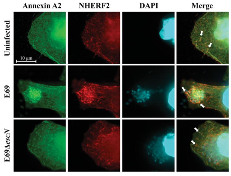
Figure 1. AnxA2 and NHERF2 co-localize in human cultured cells.
Fluorescence microscopy of HeLa-NHERF2 cells transfected with GFP–AnxA2, uninfected or infected with E2348/69 (E69) or E2348/69ΔescN strains. A total of 100 transfected cells were examined in triplicate. The green GFP signal corresponds to AnxA2, NHERF2 was stained with mouse anti-HA followed by Cy3-conjugated anti-mouse antibodies (red) and DNA from bacteria and eukaryotic cells was stained with DAPI (cyan). White arrows indicate partial co-localization of AnxA2 and NHERF2 in uninfected or E2348/69ΔescN-infected cells and intensified co-localization upon infection of E2348/69.

