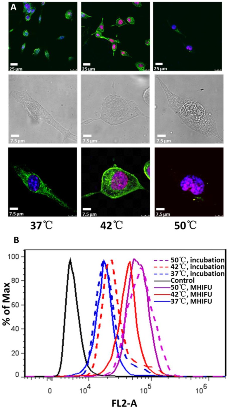
Figure 4.
Qualitative and quantitative analysis of cellular uptake at various temperatures by confocal microscope and flow cytometry. (A) The fluorescence image shows the drug has entered into the cellular nuclei after MHIFU irradiation at 42 °C while the nuclear fragmentation and myofilament destruction were occurred at 50°C. (B) Flow cytometry result shows the cellular uptake increases with the temperature elevation and cellular uptake irradiated by MHIFU at 42°C was 1.5 times larger than that of incubation.

