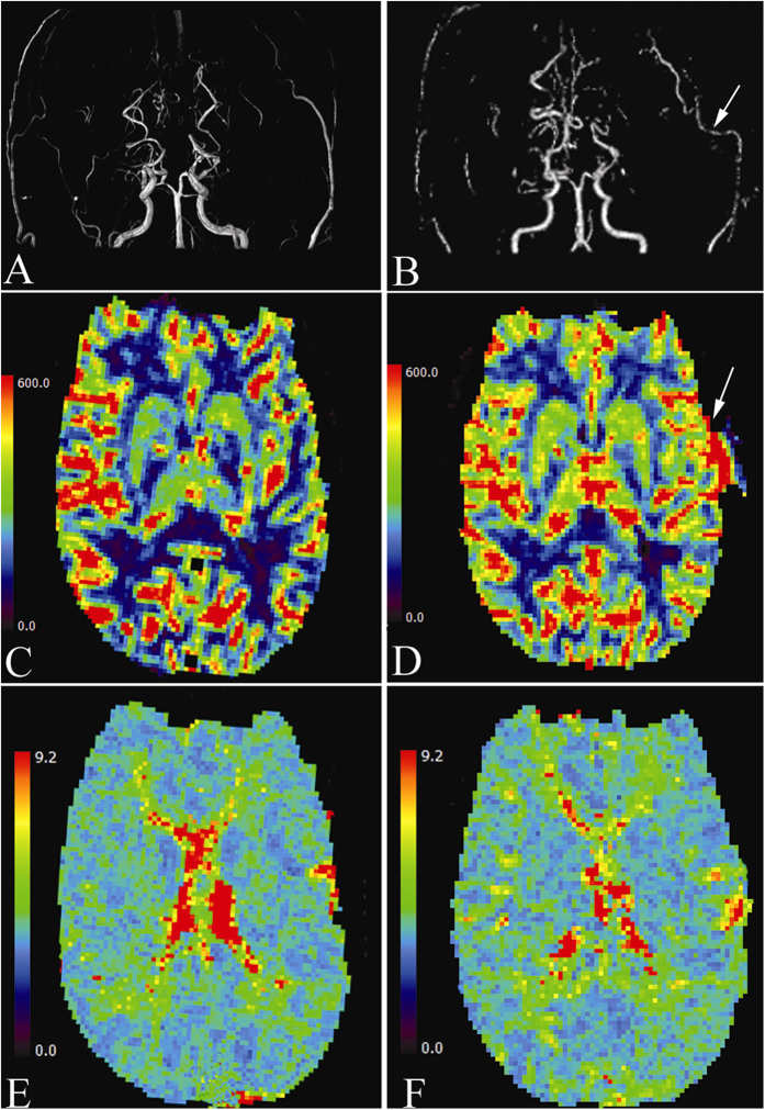
Figure 3.
(A) Preoperative MRA showing bilateral occlusion of the proximal ICA and Moyamoya change of intracranial vessels. (B) Intraoperative MRA showing the patency of left STA-MCA bypass graft (arrow). (C) Preoperative PWI demonstrating diminished CBF of the left hemisphere. (D) Intraoperative PWI demonstrating CBF of the left hemisphere markedly increased, mainly at the terminal MCA territory (arrow). (E) Preoperative PWI demonstrating prolonged MTT of the left hemisphere, especially at the terminal MCA territory. (F) Intraoperative PWI indicating the prolonged MTT of the left hemisphere didn’t change obviously.

