Figure 8.
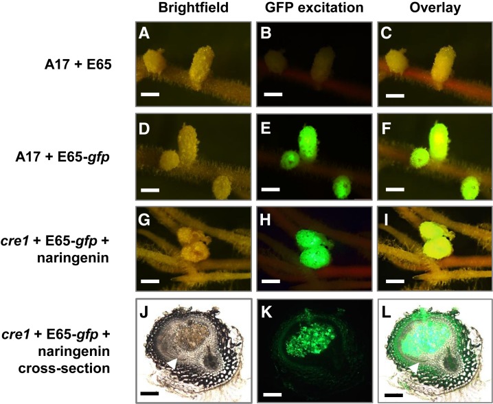
Nodules Restored on cre1 Mutant Roots Treated with Selected Flavonoids Are Infected by Rhizobia.
Nodules were quantified 3 weeks after treatment.
(A) to (C) Wild-type (A17) roots inoculated with E65 without gfp to show background autofluorescence under GFP excitation.
(D) to (F) A17 roots inoculated with a gfp-expressing Sm1021 pE65 strain.
(G) to (I) cre1 mutant roots inoculated with a gfp-expressing Sm1021 pE65 strain and treated with naringenin (3 µM).
(J) to (L) Cross section of a cre1 naringenin-rescued nodule shown in (G) to (I), with gfp-expressing Sm1021 pE65 in infected cells of the nodule.
(A), (D), (G), and (J) Bright-field images,
(B), (E), (H), and (K) Images taken under GFP excitation (maximum excitation 470 nm; 515-nm long-pass filter) of the same nodules as in (A), (D), (G), and (J).
(C), (F), (I), and (L) Overlay of bright-field and fluorescence images from the same row. More than 20 nodule-forming roots were observed under fluorescence for each treatment.
White arrowheads in (J) and (L) indicate the nodule peripheral vasculature. Bars = 1 mm in (A) to (I) and 200 µm in (J) to (L).

