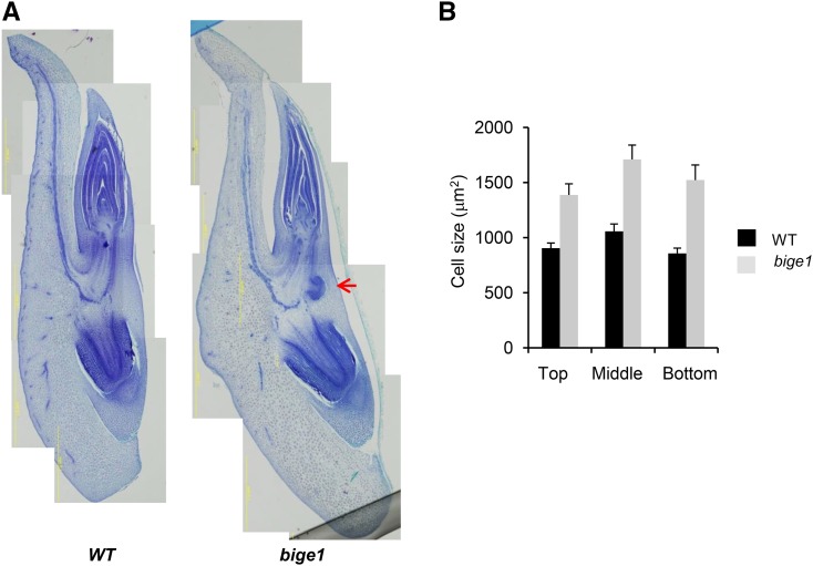
Figure 2.
Morphology of Wild-Type and bige1 Mutant Embryos.
Developing embryos of wild-type and bige1-umu1 mutant seeds were dissected from ears of self-pollinated heterozygous plants at 24 DAP.
(A) Representative paraffin thin sections prepared from wild-type and mutant embryos. The red arrow indicates a precocious lateral root primordium.
(B) Cross-sectional area of cells (n = 52 for the wild type and n = 32 for bige1) located in the apical, middle, and basal regions of the scutellum, respectively, were measured from the digital images using ImageJ (Supplemental Figure 1). The difference in cell size between the wild type and bige1 mutant was significant by Student’s t test (P < 0.03).

