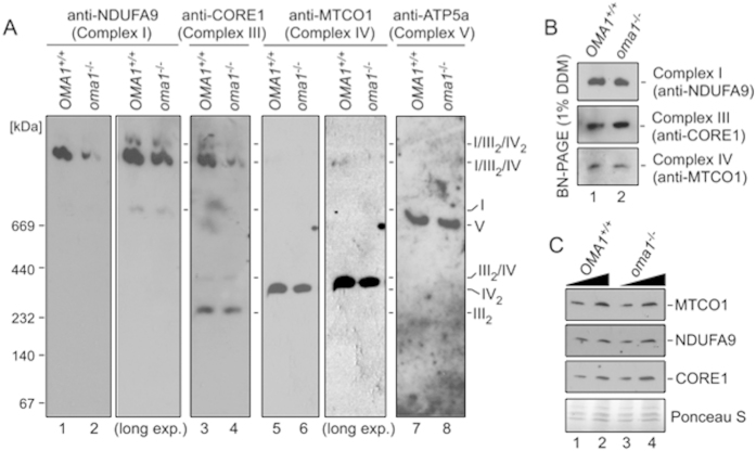
Figure 7. Loss of OMA1 impairs mammalian RSCs.
(A) BN-PAGE of mitochondria from wild type (OMA1+/+) and oma1−/− MEFs. Mitochondria (80 μg) were solubilized with 2% digitonin. Protein complexes were visualized with antibodies to NDUFA9 (Complex I), CORE1 (Complex III), MTCO1 (Complex IV) and ATP5A (Complex V). (B) BN-PAGE of WT and oma1−/− mitochondrial lysates. Mitochondria (40 μg) were solubilized with 1% dodecyl maltoside (DDM). Individual ETC complexes were visualized by immunoblotting with indicated antibodies. (C) Steady-state levels of the indicated subunits of ETC complexes in WT and oma1−/− mitochondria. Ten and 15 μg of mitochondria were analyzed by SDS-PAGE. Source data (full-length blots) for key panels of this figure are available online in Supplementary information.

