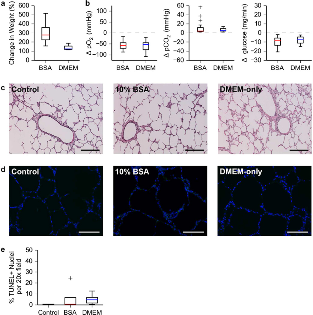
Figure 2. Bioreactor validation using short time ILC of severely damaged porcine lungs.
a. Change in organ weight after short-term (24h) ILC of porcine lungs.
b. Changes in dissolved O2, dissolved CO2, and glucose content of the culture media from the PA to the PV during short-term ILC of porcine lungs. Data shown covers three independent short-term ILCs per condition. Media was sampled 5–7 times per 24-hour period for each set of lungs cultured.
c. Hematoxylin and eosin staining of porcine lung tissue after short-term ILC. Scale bar, 250 µm.
d. Images obtained from a TUNEL assay of porcine lung tissue after short-term ILC. Nuclei and TUNEL positive cells are blue and green respectively. Scale bar, 150 µm.
e. Quantification of TUNEL positive cells in porcine lung tissue after short-term ILC.

