. 2015 Aug 24;4:e08088. doi: 10.7554/eLife.08088

Figure 2. Inhibiting Csk reduces the threshold for TCR activation and prolongs signaling induced by pMHC engagement.

(A) Purified CskAS;OTII CD4+ T cells stimulated for 3 min with 5 μg/ml bead-bound control pMHC tetramer or 5 μg/ml, 2.5 μg/ml or 1.25 μg/ml bead-bound OVA pMHC tetramer in the presence of DMSO or 10 μM 3-IB-PP1 were analyzed by immunoblotting for the phosphorylated ZAP-70, LAT and PLC-γ1, as well as total actin (loading control). Data are representative of three independent experiments. (B) Purified CskAS;OTII CD4+ T cells stimulated with 5 μg/ml bead-bound control-pMHC tetramer or 2.5 μg/ml bead-bound OVA pMHC tetramer for 2, 5 or 10 min in the presence of DMSO or 5 μM 3-IB-PP1 were analyzed for phosphorylated ERK (p-ERK) by phosphoflow. Histograms were gated on CD4+ Vα2+ cells. Data are representative of three independent experiments.

DOI: http://dx.doi.org/10.7554/eLife.08088.005

Figure 2.

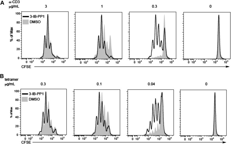
Figure 2—figure supplement 1. Csk inhibition shifts the threshold for cellular proliferation in response to anti-CD3 or p-MHC tetramer stimulation.

Figure 2—figure supplement 1.

(A) Purified CD4+ T cells from CskAS mice were loaded with CFSE and stimulated with 3 μg/ml, 1 μg/ml or 0.3 μg/ml plate-bound anti-CD3ε and 1 μg/ml anti-CD28 for 72 hr in the presence or absence of 5 μM 3-IB-PP1. Data are representative of three independent experiments. (B) Purified CD4+ T cells from CskAS;OT-II mice were loaded with CFSE and stimulated with 0.3 μg/ml, 0.1 μg/ml or 0.04 μg/ml plate-bound OVA pMHC tetramer and 1 μg/ml anti-CD28 for 72 hr in the presence or absence of 5 μM 3-IB-PP1. (A, B) Cells were then analyzed by flow cytometry for CFSE intensity. Histograms were gated on CD4+ cells. Data are representative of three independent experiments.