


Figure 3.
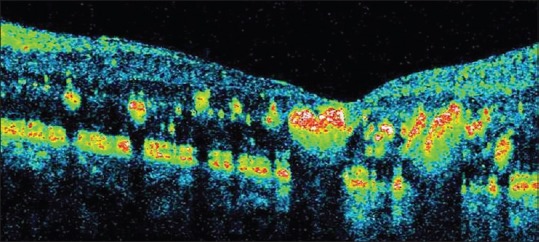
Spectral domain optical coherence tomography showing retinal thickening in areas with hard exudates and their location in outer plexiform layer.
Official websites use .gov
A
.gov website belongs to an official
government organization in the United States.
Secure .gov websites use HTTPS
A lock (
) or https:// means you've safely
connected to the .gov website. Share sensitive
information only on official, secure websites.
Spectral domain optical coherence tomography showing retinal thickening in areas with hard exudates and their location in outer plexiform layer.