
Figure 2.
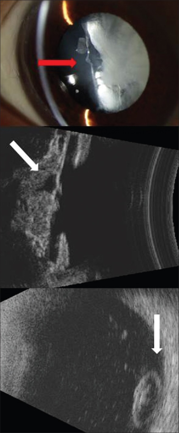
Top: Slit-lamp image showing residual cortical material within the lens capsule with absent lens nucleus and lack of typical anterior convexity (red arrow). Middle: Ultrasound biomicroscopic image showing a transpupillary section; hyperreflective cortical material within the lens capsule is visible with a hyporeflective space (oblique white arrow) representing the cavity left after the dislocation of the nucleus. Bottom: B-scan ultrasound image of the posterior segment showing a hyperreflective mass consistent with dislocated lens nucleus (vertical white arrow).