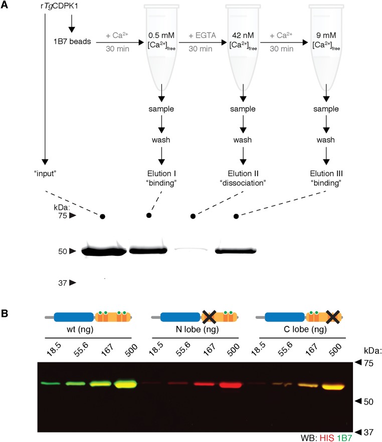
Fig. S4.
Ca2+ dependency of 1B7 binding. (A) 1B7 binds TgCDPK1 reversibly. Precipitation of recombinant TgCDPK1 by covalently immobilized 1B7 is shown. The experimental setup is diagrammed on top. Kinase was incubated with 1B7 beads, and the free [Ca2+] was modulated by adding EGTA or CaCl2, as indicated. Samples were taken after each free [Ca2+] change to determine whether complex formation had occurred, as measured by the presence of TgCDPK1 in the eluate. (B) Immunoblot comparing the detection of different kinases by IRDye800-labeled 1B7 (green). The domain structure of each kinase, indicating the KD and each of the EF hands (I–IV), is depicted for wild type (WT), D368A/D415A (N-lobe mutant), and D451A/D485A (C-lobe mutant). Detection of recombinant kinases by their His-tags (red) is included as a loading control.

