Fig. 5.
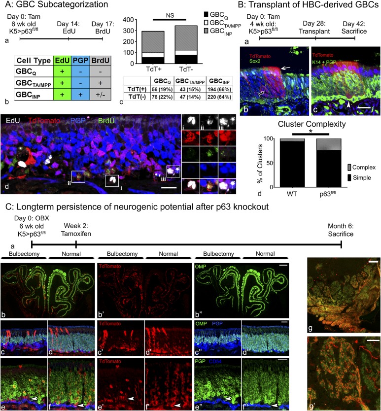
Activated HBCs generate GBCs, which integrate into the tissue and retain stem cell potential comparable to endogenous GBCs. (A) GBC subcategorization: analysis of the proliferation dynamics of endogenous TdTomato− (TdT−), and HBC-derived TdTomato+ (TdT+) GBCs. (a and b) Timeline (a) and experimental design (b) for assessing the different GBC subtypes in K5.CreERT2 > p63fl/fl;R26R(TdT) animals as described in the table. Note that the population of GBCINPs is defined by their differentiation into PGP+ sensory neurons. (c) Quantification and comparison of TdT+ (i.e., HBC-derived) and TdT− GBCs, showing no significant (NS) difference between these two GBC populations in terms of subtype distribution. (d) Representative section (included in the quantitative analysis in c) containing each of the three classifications of GBCs: (i) GBCQs: TdT+/EdU+/BrdU−/PGP−; (ii) GBCTA/MPPs: TdT+/EdU+/BrdU+/PGP−; (iii) GBCINPs: TdT+/EdU+/PGP+. (Scale bars: 10 µm in A, d; 2 µm in A, d, i–iii;) (B) Transplantation of HBC-derived cells: Transplantation of cells from K5.CreERT2 > p63fl/fl;R26R(TdT) mice [Kp63fl/flT] 4 wk after tamoxifen injection; note that the tamoxifen-treated donor animals were unlesioned at the time of harvest. (a) Timeline of experimental design. (b) An example of a complex clone consisting of morphologically distinct OSNs (open arrow) and Sox2+ Sus cells (white arrow). (c) An example of a complex clone containing a row of labeled Sus cells (arrow) and K14−/PGP− GBCs (white arrowhead). (Scale bar: 10 µm in B, b and c.) (d) Quantification of HBC-derived clones from Kp63fl/flT donors vs. WT controls (i.e., tamoxifen-treated Kp63+/+T from Fig. 1). *P < 0.05; χ2 with Yates’ correction, performed on raw data. (C) Long-term persistence of neurogenic potential after p63 knockout: Analysis of HBC-derived cells in KpT animals 6 mo after OBX. (a) Timeline of experimental design. (b–d) Mosaic image encompassing the whole section (b) and representative high-magnification images (c and d) of KpT tissue 6 mo after OBX demonstrating significant thinning of the OMP+ neuronal layer on the bulbectomized side (c) compared with the unoperated contralateral control side (d). HBC-derived TdTomato+ GBCs remain neurogenic and make a substantial contribution to the population of PGP9.5+ sensory neurons. (e and f) Representative high-magnification images from the bulbectomized side (e) and the unoperated contralateral control side (f) demonstrating the persistence of PGP9.5−/CD54− GBCs (white arrowheads). (g) On the unlesioned side, TdTomato+ OSNs (i.e., derived from recombined HBCs) actively contribute OMP+ axons to the olfactory sensory nerve, fasciculate, and extensively innervate the olfactory bulb glomeruli (g′). (Scale bars: 100 µm in C, b and g; 20 µm in C, c–f and g′.)

