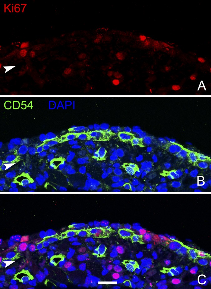
Fig. S4.
Proliferation at the time of retroviral transduction (24 hpl). Many of the Ki67+ dividing cells 24 h after MeBr lesion are CD54− GBCs situated superficial to the layer of CD54+ HBCs. (A–B) Ki67+ and CD54 staining in individual panels. (C) Overlay of all three channels to show lack of staining colocalization. Arrowheads mark the basal lamina. (Scale bar: 10 µm.)

