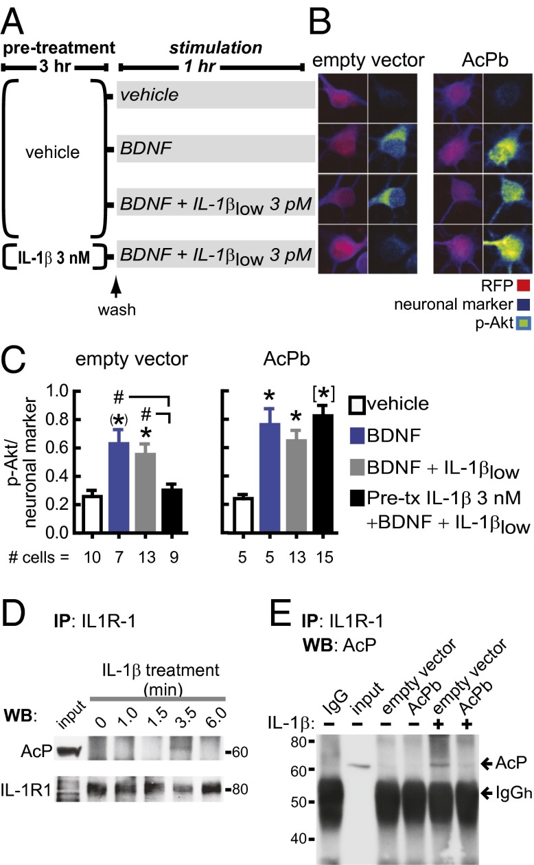
Fig. 4.
AcPb attenuates the neuronal IL-1β inflammatory response. (A) Primary rat hippocampal neurons were transfected with empty or AcPb-containing vectors at 3 DIV (AcPb gene sequence under the CMV promoter). After 3–4 d, neurons were preincubated with 3 nM IL-1β or vehicle for 3 h, washed, and stimulated with BDNF with or without IL-1βlow for 1 h, as indicated. (B) Phosphorylated levels of Akt (p-Akt; Ser-473) were assessed by immunofluorescence in RFP (reporter gene)-positive cells treated as indicated in A. Pan neuronal marker staining was used for cell-volume normalization. Representative images are shown. (Scale bar: 10 μm.) (C) p-Akt/pan-neuronal marker levels following the above treatments. The number of analyzed neurons is shown at the bottom of each treatment. Transfection itself did not interfere with BDNF signaling. Akt activation by BDNF was not impaired by IL-1βlow treatment alone (i.e., in the absence of IL-1β pretreatment) in either control- or AcPb-transfected neurons. Consistent with experiments in nontransfected cells (Fig. 2D), BDNF induction of p-Akt was prevented by IL-1βlow challenge after IL-1β pretreatment in control-transfected neurons. Bar graphs show data from a representative experiment (one out of four independent experiments). *P < 0.05; (*)P < 0.01; [*]P < 0.001 vs. control; #P < 0.05 (ANOVA, Tukey’s post hoc test). (D) Neurons were treated with 3 nM IL-1β for the indicated times. IL-1R1 immunoprecipitation (IP) was followed by Western blot (WB) for AcP and IL-1R1 (n = 3; three independent experiments). IL-1R1–AcP interaction was detected after 3.5 min, but not after 6 min, of IL-1β treatment, possibly due to MyD88 binding to the C-terminal domain of IL-1R1 (18), the same region recognized by the antibody used for IL-1R1 IP. (E) IL-1R1 IP was performed in neurons transfected with empty or AcPb-containing vectors. Neurons were treated with vehicle or IL-1β (3 nM, 3.5 min), as indicated. Membrane was probed for AcP; shown is a representative experiment (n = 3; three independent experiments). Data are presented as mean ± SEM.

