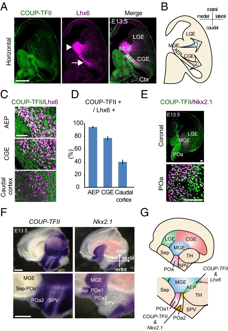
Fig. 1.
A population of the caudally migrating cells and the POa share molecular properties with the MGE and the CGE. (A) Immunohistochemical staining for COUP-TFII (green) and Lhx6 (magenta) in an E13.5 horizontal section. Multiple images were tiled and combined to show the whole brain slice. Lhx6-expressing cells form two migratory streams from the MGE (arrowhead) and the AEP (arrow). (B) Schematic diagram of the two migratory pathways. (C) Magnified views of the COUP-TFII/Lhx6–positive stream in the AEP, CGE, and caudal cortex at E13.5. (D) Quantification of the proportion of Lhx6-expressing cells that also express COUP-TFII. (E) Immunohistochemical staining for COUP-TFII (green) and Nkx2.1 (magenta) in an E13.5 coronal section, including the POa. Multiple images were tiled and combined to show the whole brain slice. High-magnification views show that COUP-TFII and Nkx2.1 are coexpressed in the ventricular zone of the POa. (F) Whole mount in situ hybridization of COUP-TFII and Nkx2.1 in the E13.5 hemisphere. COUP-TFII and Nkx2.1 are coexpressed in the POa2. (G) Schematic diagram of the expression of COUP-TFII, Nkx2.1, and Lhx6 in the POa and the AEP. Ctx, cortex; LGE, lateral ganglionic eminence; Sep, septum; SPV, supraoptic paraventricular region; TH, thalamus. [Scale bars, (A and F) 500 µm, (E) 100 µm, and (C) 50 µm.]

