Fig. 6.
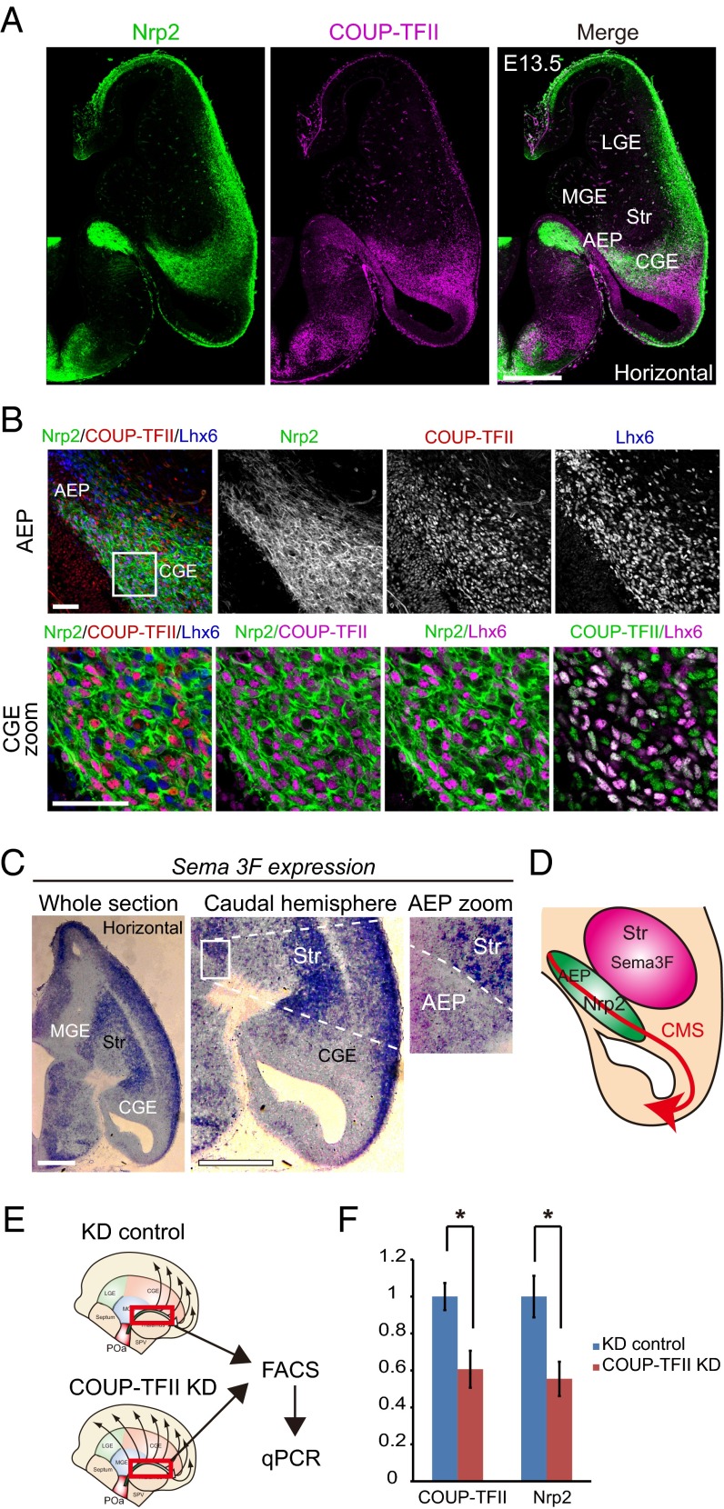
Nrp2 is preferentially expressed in the AEP and the CGE and is regulated by COUP-TFII. (A) Immunohistochemical staining for Nrp2-tauGFP (green), COUP-TFII (magenta) and Lhx6 (blue) in an E13.5 horizontal section of a heterozygous Nrp2-Δ mouse brain (+/−). Multiple images were tiled and combined to show the whole brain slice. Whole section micrographs of Nrp2 and COUP-TFII show that Nrp2 is preferentially expressed in the AEP and CGE. (B) Magnified views of the region of Nrp2/COUP-TFII/Lhx6 expression in the AEP and the CGE. Virtually all COUP-TFII–expressing cells in the CGE also express Nrp2. (C) In situ hybridization of Sema3F in an E13.5 horizontal section. Sema3F is highly expressed in the striatum region (Str) and weakly expressed in the CMS. A magnified image shows the Sema3F expression contrast between the AEP and striatum. (D) Schematic diagram of Nrp2 and Sema3F expression in the CMS. (E) Schematic representation of the experimental design for real-time PCR. POa-derived cells migrating into the CMS (red squared) were dissected from the electroporated brain, sorted by FACS, and analyzed by quantitative PCR (qPCR). (F) qPCR results following COUP-TFII knockdown. Transfection of shRNA-COUP-TFII significantly reduced the endogenous expression level of COUP-TFII and Nrp2 mRNA in migrating POa-derived cells (COUP-TFII, *P = 0.021; Nrp2, *P = 0.016; two-tailed Student’s t test). Str, striatum. [Scale bars, (A) 500 µm, (B) 50 µm, and (C) 500 µm.]

