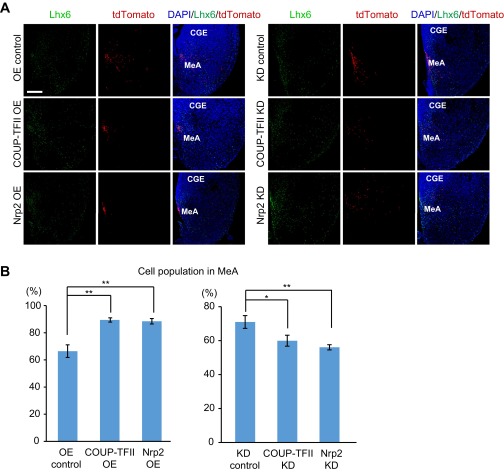
Fig. S8.
Expression levels of COUP-TFII and Nrp2 affect the distribution of POa-derived cells into the medial amygdala. (A) Cell distribution of the POa-derived cells electroporated at E11.5 was examined in coronal sections of the medial amygdala at E13.5. Immunohistochemistry for Lhx6 was performed to visualize the dorsal part of the medial amygdala. When COUP-TFII or Nrp2 was OE, the POa-derived cells labeled with tdTomato at E11.5 accumulated in the medial part of the region with Lhx6-expressing cells. In contrast, the POa-derived cells sparsely distributed when COUP-TFII or Nrp2 was knocked down (KD). (B) Proportions of tdTomato-positive POa-derived cells in medial amygdala among all tdTomato-positive cells on each coronal section were calculated. Asterisks indicate a significant difference between the control and the OE or KD samples (*P < 0.05, **P < 0.01; COUP-TFII OE, P = 0.0002; Nrp2 OE, P = 0.0003; COUP-TFII KD, P = 0.036; Nrp2 KD, P = 0.006, six brains each, Dunnett’s test). CGE, caudal ganglionic eminence; MeA, medial amygdala. (Scale bars, 100 µm.)

