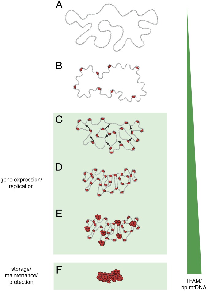
Fig. 2.
Model for packaging mtDNA into the mitochondrial nucleoid. (A) Outline of the naked mtDNA duplex (gray). (B) TFAM molecules (red) bind to mtDNA and induce bending. (C) TFAM bridges neighboring mtDNA duplexes (arrows) by cross-strand binding. (D) A combination of mtDNA duplex bending and cross-strand binding by TFAM compacts mtDNA. (E) Further compaction of mtDNA by cooperative TFAM binding into patches. (F) The final tightly packaged mtDNA in the mitochondrial nucleoid. The green triangle illustrates increasing concentration of TFAM protein per base pair mtDNA.

