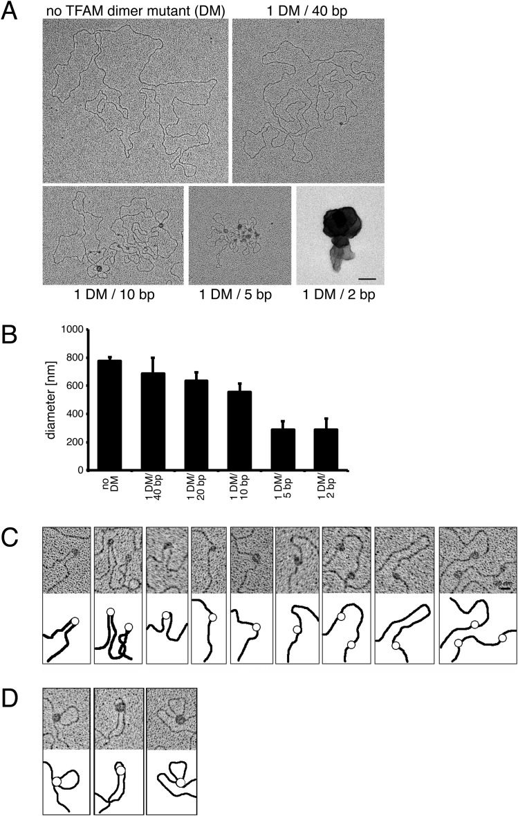
Fig. S2.
(A) Electron micrographs of spread DNA incubated with increasing concentrations of TFAM dimer mutant (DM). (Scale bar: 100 nm.) (B) Quantification of the diameters of DNA incubated with increasing concentrations of TFAM DM protein by the mean of the long and short axis. Data are represented as mean ± SD; n = 82. (C and D) Electron micrographs showing that TFAM DM binds to DNA in two different ways. TFAM binds single DNA duplexes as beads on a string inducing bending of DNA (C) or bridges two DNA duplexes resulting in loops (D). (Scale bar: 20 nm.)

