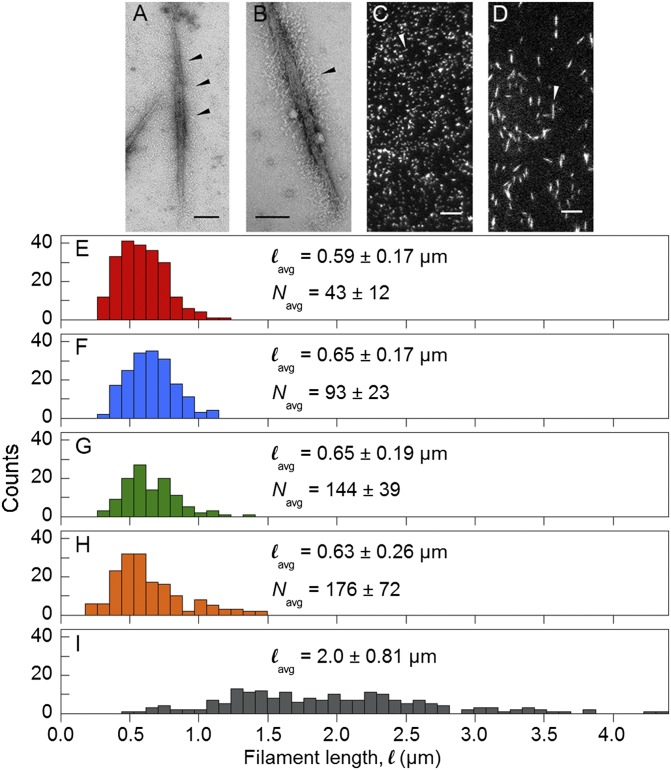
Fig. 2.
Characterization of filament preparations. (A and B) Electron micrographs of negatively stained rhodamine-labeled and EDC cross-linked 100% SMM filaments from ref. 21 and 26% SMM cofilaments, respectively. (Scale bar, 100 nm.) Arrow heads point to S1/S2 regions projecting from filament backbone. (C and D) TIRF microscopy images of rhodamine-labeled, EDC cross-linked 100% SMM filaments from ref. 21 corresponding to H and 100% SMM filaments corresponding to I, respectively. (Scale bar, 5 µm.) Arrow heads point to individual filaments. (E–I) Length distributions of 26% SMM cofilaments (E, red), 51% SMM cofilaments (F, blue); 75% SMM cofilaments (G, green); 100% SMM filaments (H, orange); and variable length 100% SMM filaments (I, gray). Colors match points in Figs. 3 and 5. lavg and Navg ± SD are indicated.

