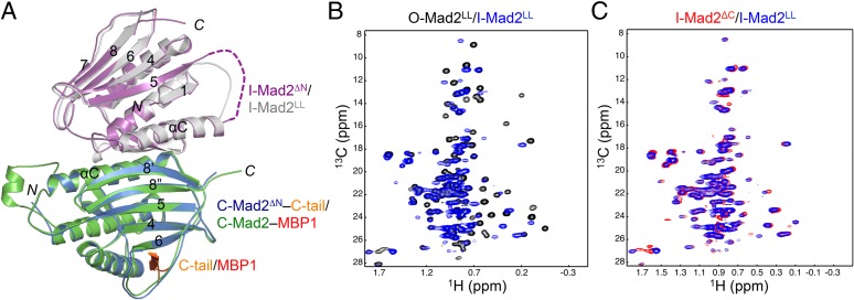
Fig. 2.
The loop-less Mad2 (Mad2LL) forms the I-Mad2–C-Mad2 dimer. (A) Superimposed diagrams of the Mad2ΔN dimer (with its I-Mad2 and C-Mad2 monomers colored in purple and blue, respectively) and the Mad2LL dimer (with its I-Mad2 and C-Mad2–MBP1 monomers colored gray and green, respectively). C-tail, the C-terminal tail of Mad2; MBP1, Mad2 binding peptide 1, an unnatural peptide ligand of Mad2 identified through phage display. (B) Overlay of the methyl region of the 1H/13C HSQC spectra of 13C-labeled O-Mad2LL (black) and 13C-labeled I-Mad2LL bound to unlabeled C-Mad2–MBP1 complex (blue). (C) Overlay of the methyl region of the 1H/13C HSQC spectra of 13C-labeled I-Mad2ΔC bound to unlabeled C-Mad2–MBP1 complex (red) and 13C-labeled I-Mad2LL bound to unlabeled C-Mad2–MBP1 complex (blue).

