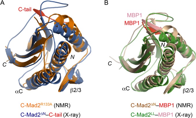
Fig. S4.
The solution and crystal structures of C-Mad2 are highly similar. (A) Superimposed diagrams of the solution structure of monomeric C-Mad2R133A (PBD ID code 1S2H; orange) and the crystal structure of C-Mad2ΔN in this study (PBD ID code 3GMH; blue). (B) Superimposed diagrams of the solution structure of Mad2∆N–MBP1 (PBD ID code 1KLQ; with Mad2 colored wheat and MBP1 colored red) and the crystal structure of C-Mad2LL–MBP1 in the Mad2LL asymmetric dimer (PBD ID code 2V64; with Mad2 colored green and MBP1 colored salmon).

