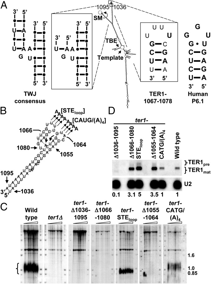
Fig. 1.
In vivo analysis of the STE region of TER1. (A) Model for the TER1 secondary structure (6). The TWJ and TER1-1067–1078 regions are enlarged to show detail. Nucleotides and base pairs that are identical to the TWJ consensus (11) or the human P6.1 sequence (13, 18) are in bold. SM, Sm binding site (6, 19); TBE, template boundary element (6, 7); Template, templating sequence; TWJ, three way junction. (B) TER1-1036–1095 enlarged. Mutants made for this study are indicated. (C) Telomere blots. Genomic DNA extracted from successively streaked spore products was digested with EcoRI and hybridized to a telomeric probe. Telomere signal is indicated by brackets. Molecular weight markers are in kb. (D) Northern blot. Total RNA extracted from ter1 mutants described in C were hybridized to TER1 and U2 probes. TER1 precursor and mature forms are indicated. Relative levels of TER1 normalized to the U2 signal are indicated below the blot.

