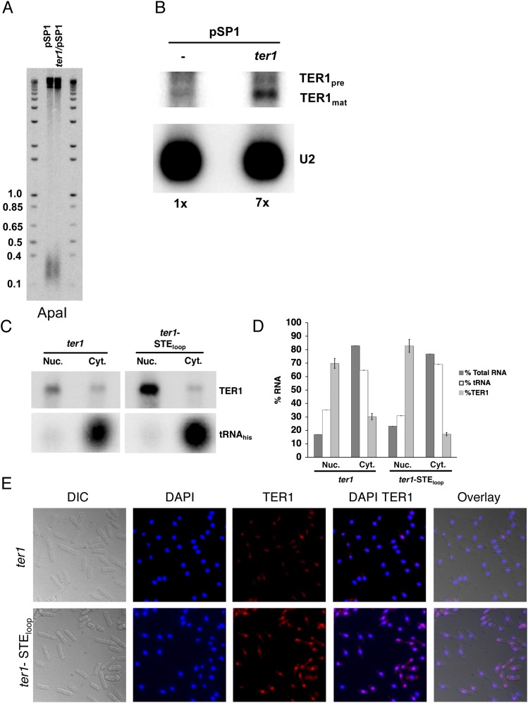
Fig. S2.
TER1 overexpression does not cause short telomeres. (A) Southern blots. Genomic DNA was extracted from cells harboring pSP1 and ter1/pSP1 after seven successive streaks. DNA was digested with ApaI and hybridized to a telomeric probe. Molecular weight markers are in kb. (B) Northern blot. Total RNA was extracted from wild-type cells containing either pSP1 vector or pSP1 carrying ter1 expressed from its own promoter [ter1/pSP1 (6)]. Northern blotting was performed as described in Fig. 1D. Relative levels of TER1 normalized to the U2 signal are indicated below the blot. (C) TER1 and TER1-STEloop RNAs localize to the nucleus. Northern blot of RNAs isolated from cytoplasmic and nuclear fractions. TER1 was identified by hybridization as in Fig. 1D. Effective fractionation was demonstrated by localization of the tRNAhis to the cytoplasmic fraction. (D) Nuclear and cytoplasmic RNAs analyzed in C were used as substrates for quantitative RT-PCR analysis. RNAs detected in each fraction are expressed as percent of the nuclear plus cytoplasmic RNAs. Error bars are SDs. The amounts of total fractionated RNA were determined by Nanodrop spectrophotometer. (E) FISH. Cells expressing TER1 or TER1-STEloop were fixed and hybridized with TER1 (Quasar 670) fluorescence probes (Biosearch Technologies). Nuclear DNA is indicated by DAPI staining.

