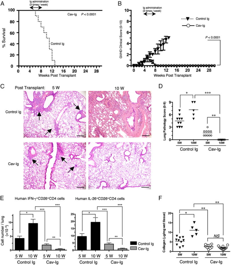
FIGURE 6.

Administration of Cav-Ig during early GVHD development impedes lethal GVHD by reducing level of IL-26+CD26+CD4 cells and collagen deposition in the lung. Sublethally irradiated NOG mice were transplanted with 1 × 10⁷ MNCs isolated from HuCB. Cav-Ig or control Ig (each 100 mg/dose) was administered i.p. thrice per week, beginning at day +29 after transplantation until day +56. (A) Overall survival and (B) clinical GVHD score (mean ± SEM). Data are cumulative results from three independent experiments (for each, n = 10). (C) H&E staining of the lung tissues of control Ig group and Cav-Ig group at 5 or 10 wk posttransplantation. Representative histology is shown from three independent experiments (for each, n = 10 in Cav-Ig group, and n = 10 at 5 wk and n = 5 at 10 wk in control Ig group). Arrows indicate perivascular and peribronchial inflammation of the small airway. Original magnification ×100. Scale bars, 100 μm. (D) Pathologic damage in the lung of recipients administered with Cav-Ig or control Ig was examined at 5 and 10 wk posttransplantation using a semiquantitative scoring system. Each dot indicates individual value, and horizontal bars indicate mean value. Data are cumulative results from three independent experiments (for each, n = 10 in Cav-Ig group, and n = 10 at 5 wk and n = 5 at 10 wk in control Ig group). *p < 0.01, **,***p < 0.0001. (E) Absolute cell number of human IFN-γ+ or IL-26+CD26+CD4 cells in the lung of recipients of Cav-Ig or control Ig group. Data are cumulative results from three independent experiments (for each, n = 10 in Cav-Ig group, and n = 10 at 5 wk and n = 5 at 10 wk in control Ig group). *,**,***p < 0.0001. (F) Collagen contents in the lung of recipients of Cav-Ig or control Ig group (5 and 10 wk posttransplantation) were quantified by Sircol Collagen Assay. The mean number (±SEM) of total collagen contents (μg) per wet lung tissue weight (mg) was determined from three independent experiments (for each, n = 10 in Cav-Ig group, and n = 10 at 5 wk and n = 5 at 10 wk in control Ig group). Each dot indicates individual value, and horizontal bars indicate mean value. *,**p < 0.0001.
