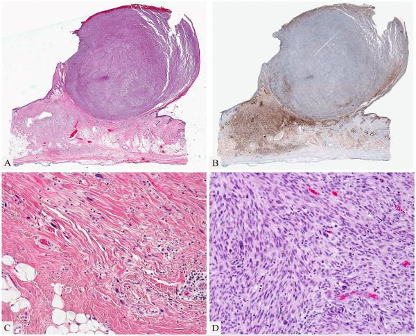
Figure 3.
A spindle cell nodule associated with desmoplastic melanoma. Spindle cell nodule protruding above the skin surface and adjacent fibrosing process in dermis and subcutis (A). Desmoplastic melanoma is highlighted by an immunostain for S100 protein while the cellular spindle cell nodule is negative for S100 protein (B). The desmoplastic melanoma shows a pauci-cellular infiltrate of hyperchromatic spindle cells in a fibrous stroma (C). The large nodule is composed of densely cellular pleomorphic spindle cells (D).

