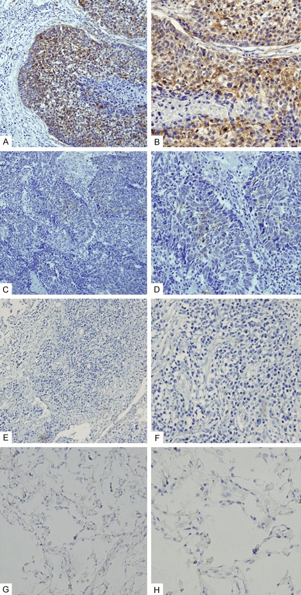
Figure 1.

Expression of Vav1 in human NSCLC specimens. A, B. High-intensity cytoplasmic staining of Vav1 in NSCLC specimens. C, D. Low-intensity cytoplasmic staining of Vav1 in NSCLC specimens. E, F. Negative immunoreactivity of Vav1 in NSCLC specimens. G, H. Vav1 expression was not observed in normal lung tissues. A, C, E, G ×200; B, D, F, H ×400.
